
Wednesday, October 29, 2003
3815
P59: Orbital Anthropometrics after Medial Canthopexy with Ipsilateral Titanium Microanchor Device
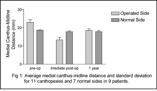
Management of medial canthal deformities still remains a great challenge in craniofacial surgery. Current techniques of transnasal canthoplasty and canthopexy present an elevated rate of relapse and malpositioning. Also, the risk of lesion to the contralateral orbit is considerable. Ipsilateral techniques used in the past did not show, as well, satisfactory results. The aim of the present study was to evaluate an alternative method for repair of medial canthal tendon displacement, using a microanchor system (1.3mm Micro QuickAnchor®, Mitek® Products, Westwood, MA, USA). Nine patients with medial canthal dystopia, seven unilateral and two bilateral, secondary to trauma, tumor resection or congenital craniofacial deformities were treated with this approach. Thecnically, the microanchor system was of easy instrumentation, allowing a good positioning of the medial canthus and avoiding transnasal drilling. Pre-operative, immediate post-operative and 1 year follow-up anthopometrics of the orbital region were evaluated, including measurement of intercanthal distance , medial canthus-midline distance, lateral canthus-midline distance and orbital width. After an initial overcorrection, the medial canthus tended to reach and sustain a position similar to the normal side (Fig 1). As observed in other series, using ipsilateral or transnasal approaches, a partial relapse occurred in two patients. Stability of anchor positioning was observed in all patients. Partial relapse of medial canthal positioning was probably due to stretching or breaking of the suture-tendon interface. In conclusion, the use of michroanchor system for medial conthopexy can be considered an easy and effective option for treatment of canthal dystopia.
View Synopsis (.doc format, 87.0 kb)
See more of Posters
Back to Plastic Surgery 2003 Complete Scientific Program
Back to Plastic Surgery 2003 Meeting home