
Wednesday, October 29, 2003
3754
P53: Intrathoracic Expandable Prostheses for the Treatment of Post-Pneumonectomy Syndrome
Background
Post-pneumonectomy syndrome is a relatively rare, unusual complication of pneumonectomy resulting from excessive shift of mediastinal structures into the empty pleural space. The most common presentations are progressive exertional dyspnea, recurrent respiratory infections, and stridor. Symptoms are secondary to extrinsic tracheal or bronchial compression by herniated mediastinal structures.
Methods
We retrospectively reviewed the charts of all patients treated at the Mayo Clinic for post-pneumonectomy syndrome from 1991-2002. Specifically, we limited our review to patients treated surgically by mediastinal repositioning utilizing intrathoracic placement of an expandable prosthesis. These procedures were performed collaboratively by the thoracic and plastic surgical services.
Results
Four(4) patients were identified who underwent surgical treatment of post-pneumonectomy syndrome. The average age at diagnosis was 48.5 years. All patients had undergone pneumonectomy for lung cancer. Patients had between 1 and 3 saline filled expanders placed intrathoracically. The average volume of saline used was 731 cc. All patients underwent intraoperative bronchoscopy to confirm resolution of bronchial compression. Follow-up averaged 30 months( range 5 –57). There were no deaths among our patients. No implants have ruptured nor have any of the patients required removal of any implants for other reasons during the follow-up period. All patients had clinical, bronchoscopic, and radiologic evidence of resolution of symptoms and signs of post-pneumonectomy syndrome.
Conclusions
Post-pneumonectomy syndrome is a rare but devastating complication following thoracic surgery. The use of intrathoracic saline filled tissue expanders alone or in conjunction with thoracoplasty provides a simple means of restoring intrathoracic volume and improved pulmonary function. Complications were few and overall survival was excellent in our patient population
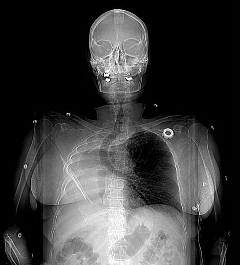
Figure1: Preoperative CT Chest scout film showing significant mediastinal shift to the right with noticeable tracheal deviation
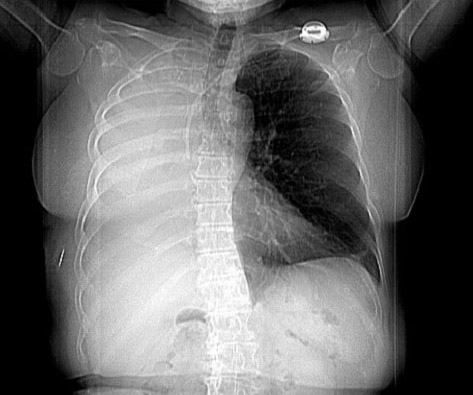
Figure 2: Postoperative CT Chest scout film after placement of intrathoracic tissue expander
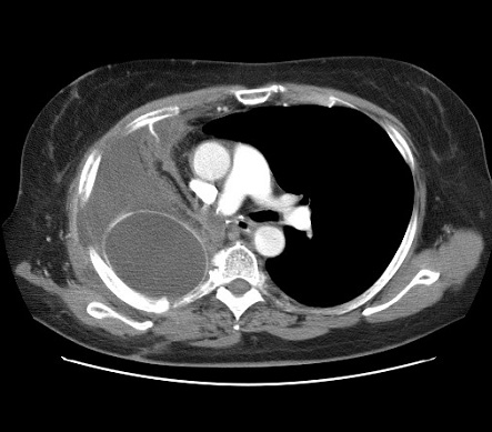
Figure 3: CT chest after placement of intrathoracic tissue expander. Mediastinal structures, trachea, and left main stem bronchus shifted towards midline.
View Synopsis (.doc format, 364.0 kb)
See more of Posters
Back to Plastic Surgery 2003 Complete Scientific Program
Back to Plastic Surgery 2003 Meeting home