
Wednesday, October 29, 2003
3752
P04: Capacity of Old Chondrocytes to Engineer Neocartilage in Vivo
INTRODUCTION: Elderly patients may require cartilage repair using old autologous chondrocytes. Most studies on tissue engineering cartilage used chondrocytes from post-fetal and very young animals. We hypothesize that articular chondrocytes from old animals produce neocartilage as well as young in vivo.
METHODS: Articular cartilage was harvested from 3-6 month old sheep (“young”) (n=5), and 8 year-old sheep (“old”) (n=4). Old and young cell were isolated separately, and mixed with fibrin glue polymer at a concentration of 40X106 cells/cc. Cell-polymer constructs were implanted into the subcutaneous tissue of nude mouse, and harvested after 7 and 12 weeks. Constructs and native cartilage (control) were assayed for H&E, and Safranin-O staining; total DNA, glycosaminoglycan (GAG), and hydroxyproline (total collagen) content.
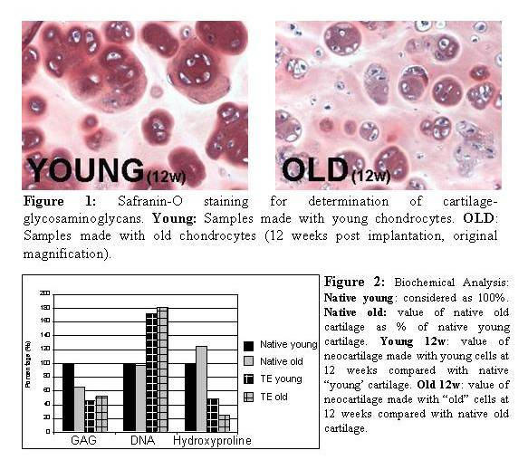
RESULTS: Samples made with both old and young chondrocytes evidenced cartilage extracellular matrix formation with H&E and Safranin-O staining at 7 and 12 weeks (Figure 1). Bioanalysis of constructs revealed that old chondrocytes increased the DNA (cell number), GAG, and Hydroxyproline content over time in a similar pattern to young chondrocytes (Fig. 2). At 12 weeks GAG content reached 80% of native old cartilage values, while hydroxyproline content reached 21%.
CONCLUSION: This study demonstrates that old articular chondrocytes can produce tissue engineered cartilage in vivo. Old chondrocytes formed cartilage-like extracellular matrix similar to that of young chondrocytes when suspended in fibrin glue scaffold. This data suggests that the engineering of neocartilage using autologous chondrocytes from elderly donors may be a feasible option for aesthetic and reconstructive procedures in elderly patients.
This study was supported by the Plastic Surgery Educational and Research Foundation
View Synopsis (.doc format, 229.0 kb)
See more of Posters
Back to Plastic Surgery 2003 Complete Scientific Program
Back to Plastic Surgery 2003 Meeting home